Posts

Coronary artery disease

Image
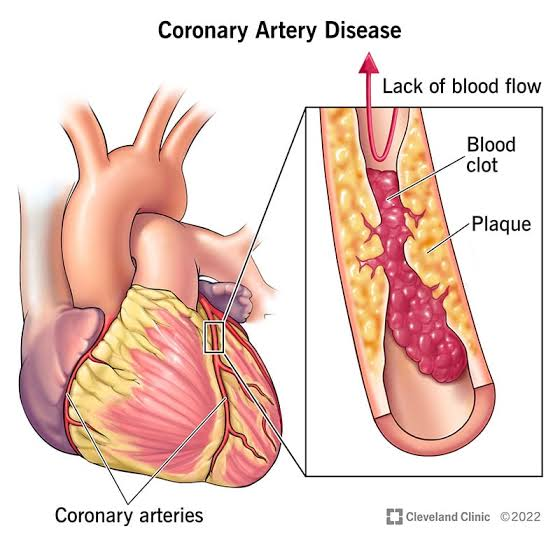
 CORONARY ARTERY DISEASE (CAD) The commonest form of coronary artery disease is narrowing of the vessel lumen by atherosclerotic plaque(s).The most frequent symptom of CAD is chest pain (angina pectoris) due to myocardial ischemia. Coronary Arteries  There are two main coronary arteries which branch to supply the entire heart. They are named the left and right coronary arteries,and arise from the left and right aortic sinuses within the aorta. Branches of Left coronary artery are: 1. Left anterior descending artery (LAD) or anterior interventricular artery 2. Left marginal artery (LMA) 3. Left circumflex artery (Cx) Branches of Right coronary artery are: 1. Right Marginal artery 2. Posterior interventricular artery MYOCARDIAL ISCHEMIA Ischemia results in immediate changes which can be detected by echo: Abnormalities of wall motion (hypokinetic, akinetic, dyskinetic) Abnormalities of wall Thickening Abnormalities of overall LV function (e.g. EF) These can be detected by 2-D echo but M-

KUB Ultrasound

Image
Ultrasound of the Kidneys In medicine, KUB refers to a diagnostic medical imaging technique of the abdomen and stands for Kidneys, Ureters, and Bladder, although in fact the Ureters only show if they are abnormally distended.  A KUB ultrasound is an examination requested by your doctor to evaluate the urinary tract (which includes the kidneys, ureters, and urinary bladder). In the male patient, the prostate gland is also scanned. A KUB Ultrasound may be requested: 1/To look for changes in the bladder wall  2/To look for changes in the kidney size or structure 3/To look for stones in the urinary tract  4/To evaluate reasons why you have recurrent kidney infection 5/To identify the cause of renal or pelvic pain  KUB Ultrasound:- The urinary bladder can only be properly assessed when full or distended, as bladder volume measurements are taken whilst the bladder is full.   The bladder and both kidneys are also scanned after the bladder has been emptied to evaluate the volume of urine ret

Deep vein thrombosis

Image
 Deep vein thrombosis  DVT stands for Deep Vein Thrombosis. It is a medical condition where a blood clot forms in one of the deep veins in the body, usually in the leg. This can cause pain and swelling in the affected area, and if left untreated, can lead to more serious health problems. In DVT, a blood clot forms in one of the deep veins in the body, usually in the leg. This can cause pain and swelling in the affected area. If the blood clot breaks off and travels through the bloodstream, it can cause a life-threatening condition called a pulmonary embolism, which occurs when the clot gets lodged in the lungs. Therefore, it is important to seek medical attention if you experience symptoms of DVT. Role of radiology in DVT Radiology plays an important role in the diagnosis and treatment of DVT. Imaging techniques such as X-rays, CT scans, and MRI scans can be used to detect blood clots and assess the severity of the condition. These imaging techniques can also be used to monitor the eff

ROLE OF CT IN EVALUATION OF ABDOMINAL MASSES

 MAIN ROLE OF CT:- An abdominal CT scan makes detailed pictures of the structures inside your belly very quickly. This test may be used to look for: Cause of blood in the urine. Cause of abdominal pain or swelling. Palpable abnormalities, such as abdominal mass or organomegaly. Both US and CT are excellent techniques for confirming or excluding the presence of an abdominal mass, with sensitivity and specificity higher than 95%. The accuracy of US in determining the organ of origin is estimated at between 88% and 91%.Mainly CT scan are 100 percent accurately in evaluation of abdominal masses. For intra-abdominal masses, contrast-enhanced CT and ultrasound examination have demonstrated accuracy. For abdominal wall masses, which may arise from muscle, subcutaneous tissue, or connective tissue, MRI, CT, and ultrasound all provide diagnostic value. An abdominal CT scan uses a special X-ray machine to take pictures of the liver, spleen, kidneys, bladder, stomach, intestines, pancreas, and ad

THE EVALUATION AND MANAGENMENT OF HEMATURIA IN SURGICAL PRACTICE.

 Hematuria means blood in urine. It is not a disease but symptom of a disease. Management of hematuria depends underlying cause of hematuria.  UTI:-          Trimethorim/sulfamethoxazole, nitrofurantoin and fosfomycin are the most preferred antibiotics for treating UTI NEPHROLITHIASIS:-                                      Kidney stones (also called renal calculi) nephrolithiasis or urolithiasis are hard deposits made of minerals and salts that form inside kidneys. Diet excess body weight, some medical conditions and certain supplements and medications are among the many causes of kidney stones.        Investigation include imaging studies eg. Xray KUB and USG abdomen+ pelvis to make a definitive diagnosis.   Management options include: 1) Shock wave lithotripsy. The doctor can use shock wave lithotripsy link to blast the kidney stone into small pieces. 2) Cyctoscopy and ureteroscopy. 3) Percutaneous nephrolithotomy BLADDER CANCER:-                             Bladder cancer is a di

Radiology and types

 Radiology is a branch of medicine that uses imaging technology to diagnose and treat various disease. Radiology may be divided into two different areas, diagnostic radiology and interventional radiology. Doctors who specialize in radiology are called radiologists. Diagnostic Radiology Diagnostic Radiology is a specialization that involves undertaking range of imaging procedures to obtain the images of inside body. The diagnostic radiologist then carefully interprets these images to diagnose illness and injury.  The most common types of diagnostic radiology exams include: CT scan also known as computed tomography, It is a type of imaging scan or test which can generate or create a very detailed image inside body. It is often used in radiology to diagnose and monitor wide range of conditions including cancer, heart diseases, bone fractures, brain , chest , abdomen etc.  Patient lies on a table that moves through doughnut-shaped machine that takes xray from different angles. CT scans are

Basics of ultrasound.

Image
 Ultrasound is a type of imaging that uses high-frequency sound waves to look at organs and structures inside the body. Gray scale Gray scale is a type of ultrasound image that uses different shades of gray to show the different densities of tissue within an organ or structure. M mode and B mode M mode is a type of ultrasound imaging that shows how structures within the body move over time. It is often used to look at the heart and blood flow. B mode is a type of ultrasound imaging that shows a two-dimensional image of organs and structures in the body. It is often used to look at the fetus during pregnancy, as well as other organs like the liver, kidneys, and thyroid. Roles of Ultrasound Ultrasound is a medical imaging technique that uses high-frequency sound waves to create images of organs and structures inside the body. It is non-invasive, safe, and painless. Ultrasound images are created by sending sound waves into the body and measuring the echoes that bounce back. These echoes a

Initials about Radiology

 Radiology is a medical specialty that uses imaging techniques to diagnose and treat diseases. Radiology is a field of medicine that uses imaging techniques such as X-rays, CT scans, MRIs, and ultrasounds to diagnose and treat diseases. Radiologists are medical doctors who specialize in interpreting these images to help diagnose and treat patients. They work closely with other doctors and healthcare professionals to provide the best care possible. Radiology is a rapidly evolving field that has advanced significantly in recent years. With the help of advanced imaging techniques, radiologists can now detect diseases and injuries earlier and more accurately than ever before. This has led to improved patient outcomes and a better understanding of the human body. Radiologists use a variety of imaging techniques to diagnose and treat diseases, including X-rays, CT scans, MRIs, and ultrasounds. They work closely with other healthcare professionals to provide the best possible care to their pa